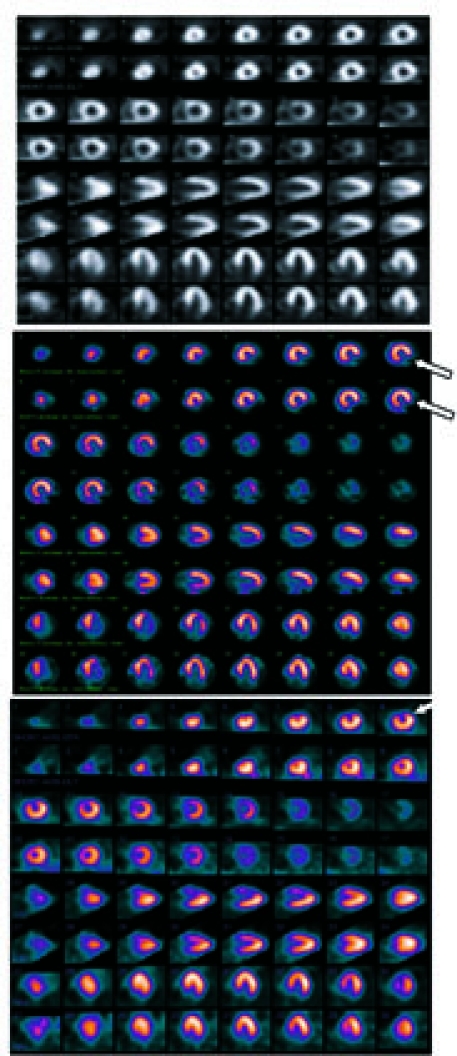
Fig. 1 Examples of myocardial perfusion images in a cancer population. The top panel (normal) shows a normal perfusion image, the middle panel (scar) indicates a fixed defect (double arrows), and the bottom panel (ischemia) indicates a reversible defect (single arrow).
