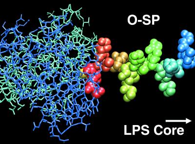
Figure 5.
Model of the Ab–O-SP complex based on the structure of the Fab–disaccharide complex. Individual perosamine residues are shown in different colors; the light and heavy chains of the Ab variable domains are shown in light and dark blue, respectively. The O-SP Ag was built assuming that all glycosidic linkages display the same dihedral angles observed for the Fab-bound disaccharide, giving rise to an extended polysaccharide conformation. The upstream terminal perosamine is bound inside the Ab-binding cavity (the 2-O-methyl group at the center of the interface is shown in yellow). As revealed by the Fab–disaccharide structure, the second perosamine residue is positioned at the exterior of the binding site and makes fewer contacts with Ab residues, whereas subsequent sugar residues are not involved in the interaction. Figure produced with the program vmd (42).

