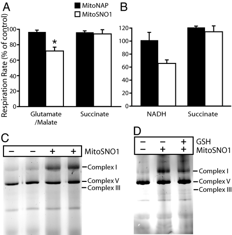
Fig. 6.
Inhibition and S-nitrosation of complex I by MitoSNO1. (A) Effect of MitoSNO1 on mitochondrial respiration. Heart mitochondria were incubated in an oxygen electrode with 5 mM glutamate and 5 mM malate, or succinate, and incubated with 10 μM MitoSNO1, 10 μM MitoNAP or carrier for 2–3 min, then 250 μM adenosine diphosphate (ADP) was added and the rate of respiration measured. Respiration +MitoSNO1 is expressed as a percentage of that with MitoNAP, and are means ± SEM from 3 mitochondrial preparations; *P < 0.05 by Student's paired t test. (B) Effect of MitoSNO1 on respiration by mitochondrial membranes. Bovine heart mitochondrial membranes were incubated in an O2 electrode with 75 μM MitoSNO1 or MitoNAP, or ethanol carrier for 5 min ± rotenone, then 1 mM NADH or 10 mM succinate was added and respiration measured. Data are expressed as respiration as a percentage of the appropriate controls and are means ± range of 2 separate experiments. (C) S-nitrosation of complex I by MitoSNO1. Bovine heart mitochondrial membranes were incubated with 75 μM MitoSNO1 or carrier for 5 min. Then protein SNOs were labeled by maleimide-Cy3 and respiratory complexes were separated by BN-PAGE and scanned for Cy3 fluorescence. The locations of respiratory complexes I, III, and V were determined by immunoblotting. (D) Effect of GSH on S-nitrosation of complex I by MitoSNO1. Bovine heart mitochondrial membranes were incubated and processed as in (C) except that after incubation ± MitoSNO1 the membranes were incubated ± 1 mM GSH for 15 min.

