
Figure 3. Cell death and inflammatory responses are inhibited by prior sleep deprivation.
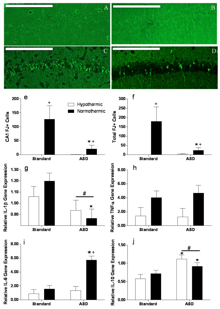
Representative Fluoro-Jade C stained sections of the CA1 field of the hippocampus following CA/CPR in A) standard housed hypothermic mice, B) ASD hypothermic mice, C) standard housed normothermic animals and D) ASD normothermic animals; scale bar = 200μm. E) Fluoro-Jade positive cells in the CA1 field and F) summed across the whole hippocampal formation. The cell death data are displayed in the figure are untransformed but statistical testing was conducted on log-transformed data. RT-PCR analyses of hippocampal mRNA 24 hours post reperfusion of the cytokines G) interleukin-1β (IL-1β), H )tumor necrosis factor alpha (TNFα), I) interleukin-6 (IL-6) and J) interleukin 10 (IL-10). + Significantly different from hypothermic mice in the same sleep condition. * Significantly different from standard housed mice. Differences were considered significant if p<0.05. ASD, acute sleep deprivation. All data are presented as means ( ± SEM). N=6–7 animal/group for histology and 5–6/group for mRNA analysis.