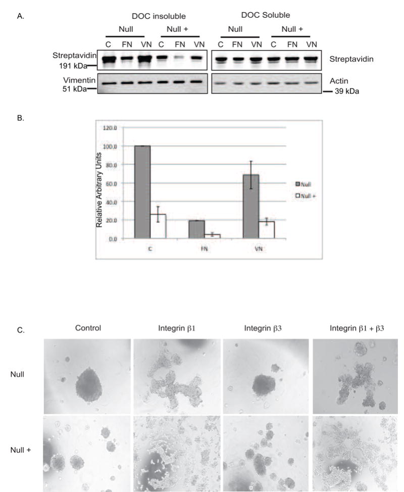
Figure 4.

Fibronectin assembly is mediated by Integrin α5β1 in Null and Null + cells: A) Exogenous assembly assay; Cells were plated on 10ug/ml fibronectin or 5 ug/ml vitronectin in fibronectin free medium prior to the addition of biotinylated fibronectin. Cells were incubated overnight followed by collection of lysates and separation into DOC-soluble and insoluble fractions. Left panels: DOC-insoluble fractions. Biotinylated fibronectin is detected by blotting with IR Dye 680 Streptavidin. Vimentin serves as a loading control. Right Panels: DOC-soluble fraction: Actin serves as a loading control. B) Densitometric analysis of DOC insoluble fraction in panel A. Gray bars represent Null cells. White bars represent Null + cells. C) Hanging drop assay: Cells were treated as in figures 1 and 2 except for treatment with 100 μg/ml inhibitory antibodies against integrins β1 (HA 2/5), β3 (2C9.G2) or a combination of both.
