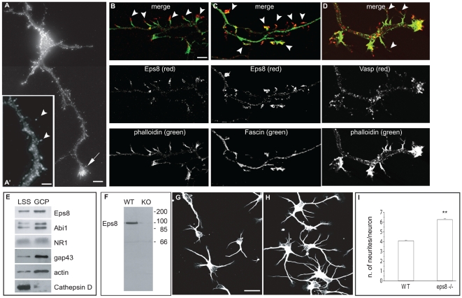
Figure 1. Eps8 is localized to sites of active actin polymerization in hippocampal neurons, and its removal increases neurite number.
(A–D) Eps8 localization in hippocampal neurons. Three days in vitro (3DIV) cultured hippocampal neurons were stained with anti-Eps8 antibody alone (A and A′) or in combination with either FITC-conjugated phalloidin to label F-actin (B), or anti-fascin (C) as indicated. The arrow in (A) indicates the axonal growth cone. In the inset (A′), the arrowheads indicate axonal protrusions (arrowheads). The arrowheads indicate Eps8- and actin- (B), or Eps8- and fascin-rich (C) filopodia protruding from the axon. (D) Actin rich axonal protrusions are also immunopositive for VASP. (E) Eps8, its binding partner Abi1, and the growth cone markers GAP43 and actin, are enriched in preparations of growth cone particles (GCPs) relative to low-speed supernatant brain homogenates (LSS). GCP preparations, described in Materials and Methods, were immunoblotted with the indicated antibodies. The immunoblot is a representative example of five independent experiments. (F) Lysates from wild-type (WT) and eps8 − /− (KO) hippocampi were immunoblotted with anti-Eps8 antibody. Molecular weight markers are indicated. (G and H) Cultured hippocampal neurons (1DIV) prepared from WT (G) or eps8 null (H) mice were fixed and stained with anti–beta-tubulin antibody. (I) Quantification of the number of neurites/neuron. Hippocampal neurons were prepared as described in (G and H). The difference between the numbers of neurites of eps8 − /− WT neurons is statistically significant (Mann-Whitney rank sum test, p≤0.001). Data are expressed as mean±standard deviation (SD); n = 897 examined WT neurons, and 760 examined eps8 KO neurons. Bars indicate 5 µm for (A, B, C, and D); 3 µm for a′; and 10 µm for (G and H). Double asterisks (**) in (F and G) indicate p<0.001.

