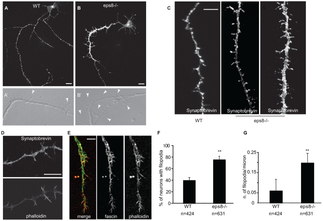
Figure 2. Genetic removal of eps8 causes increased number of axonal filopodia in hippocampal neurons.
(A and B) Cultured hippocampal neurons (3DIV) from WT and eps8 − /− mice were fixed and stained with synaptobrevin/VAMP2 antibody. The selective sorting of synaptobrevin/VAMP2 immunoreactivity to a single process reveals that only one neurite is the putative axon. Bar indicates 10 µm. (A′ and B′) Still images of phase-contrast videos of the axonal shaft of cultured hippocampal neurons (3DIV) from WT and eps8 − /− mice (see Video S1). Arrowheads highlight axonal filopodia. (C) Representative examples of axonal shafts of WT and eps8 − /− neurons, labeled with an antibody against synaptobrevin/VAMP2. Bar indicates 10 µm. (D and E) Filopodia protruding from axons were costained with anti-synaptobrevin/VAMP2 antibody and phalloidin (D) to detect synaptic vesicle and filamentous actin, respectively, or with anti-fascin and phalloidin (E), as indicated. Bar indicates 7 µm. (F and G) Neurons from eps8 − /− mice display a significantly higher density of axonal filopodia. (F) Quantification of the percentage of neurons with filopodia in WT and eps8 − /− neurons. Neurons with more than 0.04 filopodia/µm were considered as filopodia-bearing neurons (Student t-test, p≤0.001). (G) Quantification of the density of axonal filopodia in WT and eps8 null hippocampal neurons (Mann-Whitney rank sum test, p≤0.001). n = 424 examined WT neurons and 621 examined eps8 KO neurons. Data are expressed as the mean±SD.

