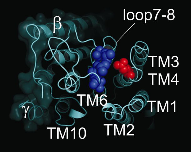
Fig. 6.
External view of the Na,K-ATPase structure[3]. Space-filling models illustrate Glutamate-309 in the M3M4 extracellular loop (red) and Leucine-879, Valine-881, and Asparagine-882 in the M7M8 extracellular loops (blue) of the pig renal enzyme. Transmembrane helices are labeled accordingly.

