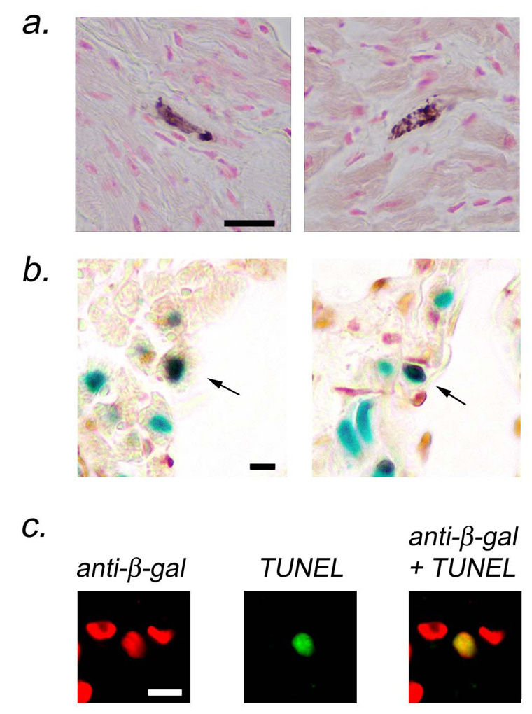
Figure 2.
Atrial cardiomyocyte apoptosis in MHC-TGFcys33 ser / MHC-nLAC double transgenic mice. (a) Activated caspase 3 immune assay for cardiomyocyte apoptosis. Cardiomyocytes at early stages of apoptosis were identified by the presence of activated caspase 3 immune reactivity (cytoplasmic brown signal in rod-shaped cells). Bar = 20 microns. (b) ISEL assay for cardiomyocyte apoptosis. Sections were stained with X-GAL to identify cardiomyocyte nuclei (blue signal) and for ISEL activity to identify apoptotic cells (HRP-conjugated reaction, dark brown signal). Apoptotic cardiomyocytes have brown and blue signals over the nucleus (arrows). Bar = 10 microns. (c) TUNEL assay for cardiomyocyte apoptosis. Sections were stained for beta-galactosidase immune reactivity to identify cardiomyocyte nuclei (rhodamine conjugated secondary antibody, red signal, left panel) and for TUNEL activity to identify apoptotic cells (FITC-conjugated secondary antibody, green signal, middle panel). Apoptotic cardiomyocytes are identified in the merged image (yellow signal, right panel). Bar = 10 microns.

