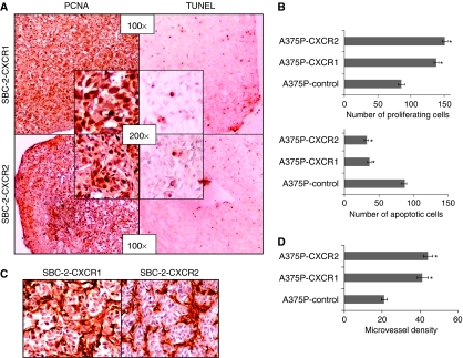
Figure 3.
CXCR1 and CXCR2 expression enhances in vivo melanoma cell proliferation, survival and tumour neovascularisation. Immunohistochemical staining for PCNA and TUNEL were analysed based on DAB staining as described in Materials and Methods. In SBC-2-group, tumours showed an imbalance of PCNA to TUNEL-positive cells (A). For A375P-group PCNA (B, upper panel) and TUNEL (B, lower panel) positive cells were counted in 10 arbitrarily selected fields at 200 × magnification in a double-blinded manner and expressed as average number of cells per field view±s.e.m. Immunohistochemical staining for microvessel using anti-GS-IB4 was analysed as described in Materials and Methods. Tumours from SBC-2 group were highly vascular (C). For A375P tumours, quantification of microvessel density at 200 × magnification in 10 random fields was examined. The values are average number of immunostained positive cells±s.e.m. (D). *Significantly different from control (P<0.05).

