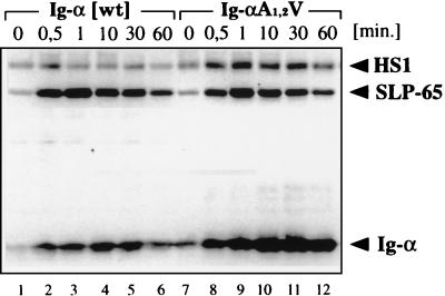
Figure 3.
The time course analysis of PTK substrate phosphorylation in stimulated J558L cells expressing either a wild-type (wt) BCR (lanes 1–6) or a BCR with an A1,2V-mutated Ig-α protein (lanes 7–12). Cells were stimulated for the indicated times with 10 μg/ml of goat anti-mouse IgM antibodies. The proteins of total cellular lysates were size-separated by SDS/10% PAGE and analyzed by 4G10 immunoblotting.

