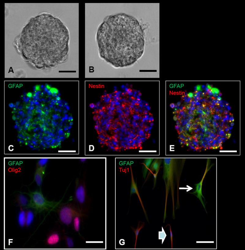
Figure 2. Neurosphere assay and differentiation from intra-operatively obtained human cells.

A, Tumor neurosphere derived from a patient with glioblastoma multiforme (GBM). B, Neurosphere derived from the subventricular zone (SVZ) in a patient who underwent a hemispherectomy for seizures. C–E, ICC images of a GBM-derived tumor neurosphere with positive GFAP (C), Nestin (D), and Nestin and GFAP co-stained (E) cells. F, Neurosphere differentiation with cells positive for GFAP (astrocytes) and Olig2 (oligodendrocytes). G, Neurosphere differentiation with cells positive for GFAP (astrocytes) and Tuj1 (neurons). Some of the cells are positive for both GFAP and Tuj1. Some of these cells have morphologies more characteristic of astrocytes with several cytoplasmic projections (arrow), while other cells are more characteristic of neurons with bipolar processes (arrowhead). This co-staining is typically present in differentiated cells from tumor-derived neurospheres, but typically not seen in differentiated cells from SVZ-derived neurospheres. Scale bars: 100μm (A–E), 10μm (F), 30μm (G).
