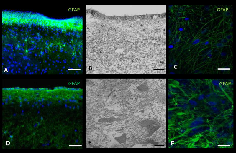
Figure 4. Immunohistochemistry (IHC) and electron microscopy (EM) of human-derived brain tumor and non-tumor tissue.

A, IHC and B, EM image of the subventricular zone (SVZ) adjacent to the body of the lateral ventricle in a non-cancer patient. C, IHC of cortical tissue in a non-cancer patient. D, IHC and E, EM image of tumor tissue in a patient with glioblastoma multiforme (GBM). F, IHC of tumor tissue in a patient with GBM. DAPI – blue. Scale Bar: A, 40μm; B, 25μm; C, 20μm; D, 20μm; E, 2μm; F, 5μm.
