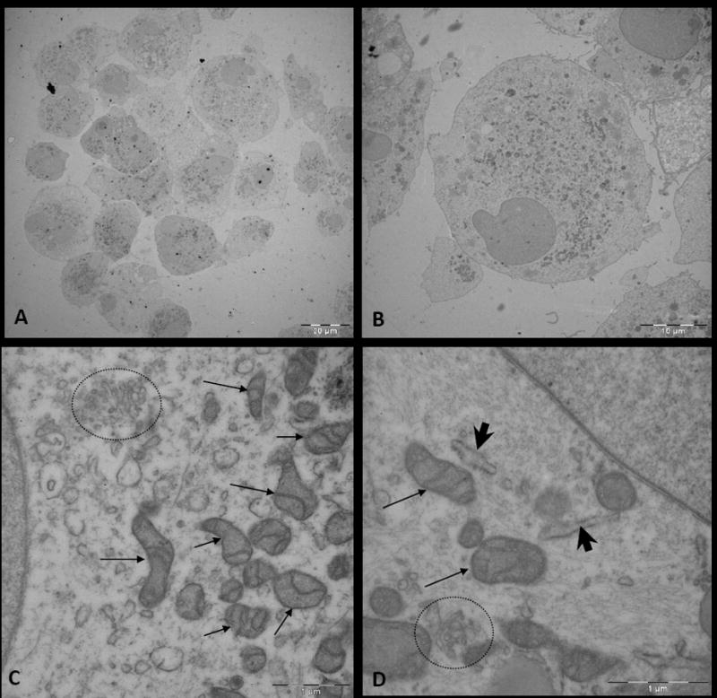
Figure 5. Electron microscopy (EM) of human-derived brain tumor neurospheres.

A, Image of cells within a glioblastoma multiforme (GBM)-derived neurosphere. B, Image of a cell within the neurosphere. C and D, Magnified images of the cytoplasm of a cell within a neurosphere, which displays abundant organelles including mitochondria (arrow), Golgi apparatus (circle), and endoplasmic reticulum (arrow head).
