Figure 5.
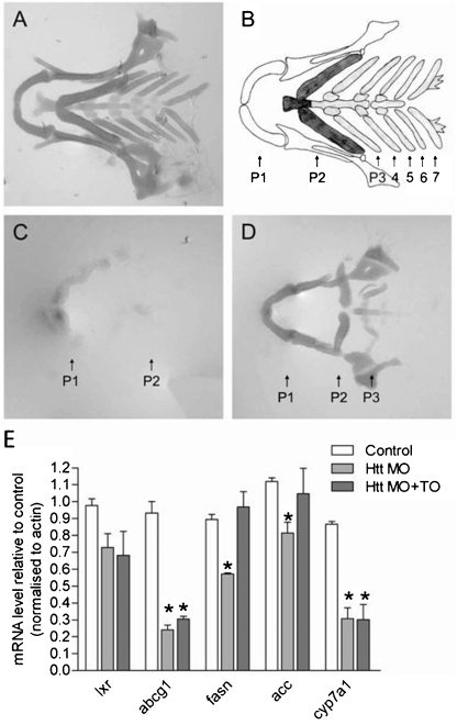
Effect of an LXR agonist on cartilage of Htt-deficient zebrafish. (A) Ventral view of an Alcian green stained lower jaw of an Htt_ATG-5mis control at 102 hpf depicting the normal zebrafish cartilage. A representative zebrafish is shown from five independent experiments where 11–22 zebrafish were analysed per experiment. (B) Diagram of the pharyngeal skeleton in ventral view showing the first or mandibular arch (P1, white), the second or hyoid arch (P2, dark grey) and five brachial arches (P3–7, light grey). (C) Cartilage is strongly reduced in Htt_ATG injected zebrafish. The mandibular (P1) and hyoid (P2) are present but small and malformed and pharyngeal arches P3–5 are missing. A representative zebrafish is shown from five independent experiments where 7–20 zebrafish were analysed per experiment. (D) An LXR agonist rescues formation (compactation, form, staining intensity) of the anterior two arches. A representative zebrafish is shown from fove independent experiments where 9–14 zebrafish were analysed per experiment. (E) Q-PCR assessment of expression of LXR and LXR target genes mRNA in uninjected control zebrafish and Htt MO injected zebrafish at 3 dpf. cDNA levels were normalized to actin levels and were plotted compared to control zebrafish (mean ±SEM, n = 3−4 experiments; *p<0.05 as analysed by one way ANOVA with Dunnett’s post test).

