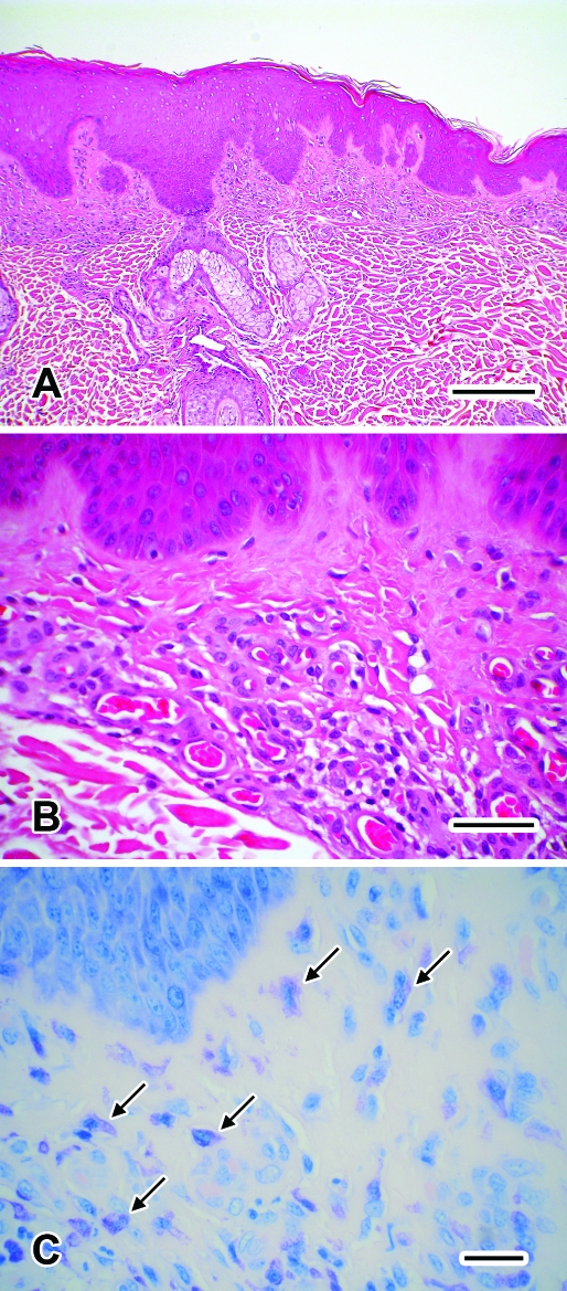
Figure 2.
Biopsy of the skin lesion from the sparsely haired-skin of the ventral mandible from the rhesus macaque from Figure 1. (A) Diffuse acanthosis of the epidermis. Hematoxylin and eosin stain; bar, 200 μm. (B) Moderate superficial dermal fibrosis and moderate thickening of the walls of superficial dermal vessels. Hematoxylin and eosin stain; bar, 50 μm. (C) Moderate numbers of mast cells (arrows) are noted with a diffuse superficial dermal distribution. Toluidine blue stain; bar, 25 μm.

