Figure 3.
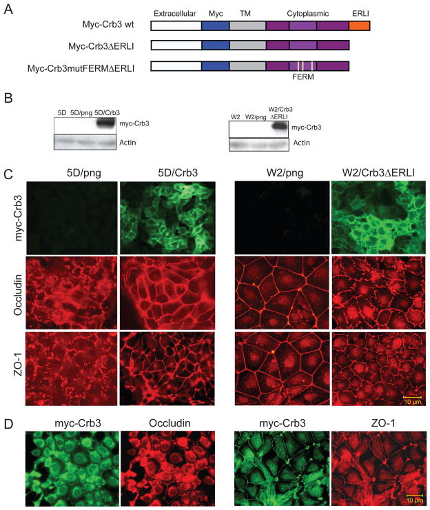
Role of Crb3 in controlling tight junction formation. (A) Graphic representation of wild type Crumbs 3 protein domain structure and schematic illustrating the Myc-tagged Crb3 and Crb3 mutants22. The Crb3 mutants contain a deletion of the C-terminal domain (ΔERLI) and three point mutations in the FERM domain (Crb3mutFERM). (B) Western blots illustrating the stable expression of myc-Crb3 in TDCL 5D and the mutant myc-Crb3ΔERLI in W2.3.1-5 cells. (C) Crb3 expression restores tight junction formation in TDCL 5D. TDCL 5D png vector infected or wild type Crb3 expressing vector infected cells and stained with for tight junction markers occludin, and ZO-1. Immunofluorescence showin restoration of tight junctions indicated by alterations in ZO-1 and occludin. Expressed Crb3 localized to the plasma membrane and rescued tight junction formation in TDCL 5D as indicated by increased occludin and ZO-1 at cell junctions compared to png vector control. Mutant crb3 expression impairs tight junction formation in parental W2.3.1-5 cells. W2.3.1-5 parental cells png vector infected or Crb3ΔERLI expressing vector infected were immunostained for tight junction markers occludin, and ZO-1. Immunofluorescence showing disruption of tight junctions indicated by alterations in ZO-1 and occludin. (D) Immunofluorescence showing co-localization of Crb3 with occludin and ZO-1 in TDCL 5DCrb3 cells.

