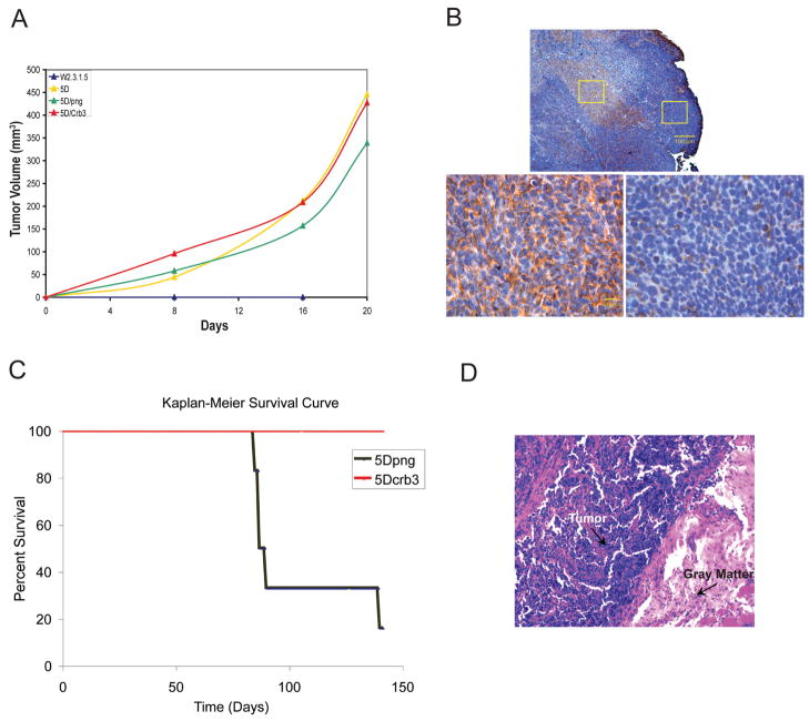
Figure 6.
Crb3 expression suppresses metastasis. (A) Tumorigenic capacity of TDCL 5D and png and Crb3 transfectants compared to parental W2.3.1-5 cells. Animals were injected subcutaneously with 107 cells and tumor formation was monitored over time. The parental W2.3.1-5 cells failed to form tumors by 20 days post injection while the TDCL 5D formed subcutaneous tumors as expected. Expression of Crb3 in TDCL 5D did not suppress subcutaneous tumor growth. (B) Subcutaneous tumor growth of TDCL 5D Crb3 is associated with the loss of Crb3 expression. TDCL 5D Crb3 tumors that formed 3 weeks post injection were examined by immunohistochemistry for Myc-Crb3 using the Myc tag. Crb3 positive cells with Crb3 localized to cellular junctions (left) were found in central (top) but not expanding, peripheral regions (right) of the tumor. (C) Crb3 expression suppresses metastasis. Kaplan-Meier survival curve of mice injected (tail vein) with 5×10⁵ cells TDCL 5D png (black, n=6) and TDCL 5D Crb3 (red, n=6). Most (5/6) of the TDCL 5D png injected animals succumbed to multiple bone and kidney metastases by 10 weeks whereas all of the TDCL 5D Crb3 injected animals remained tumor free. (D) Immunohistochemistry showing metastasis to the spinal cord, illustrated by purple cells invading the gray matter (pink area) in animals injected with TDCL 5D png cells.

