
Fig. 9.
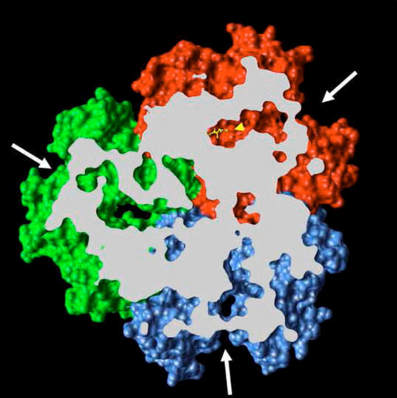
Periplasmic domain of the asymmetric crystal structure of AcrB. The view is from the top (periplasm), and the proximal portion is removed as identified by the white slab plain. The binding protomer (red) contains bound minocycline (ball-and-stick representation in yellow, identified by a yellow arrowhead). The large external cleft (white arrows) is open in the binding protomer as well as in the access protomer (blue), but is closed in the extrusion protomer (green). The figure is based on the PDB file 2DRD, and was drawn by using UCSF Chimera package [69] from the Resource for Biocomputing, Visualization, and Informatics at the University of California, San Francisco.