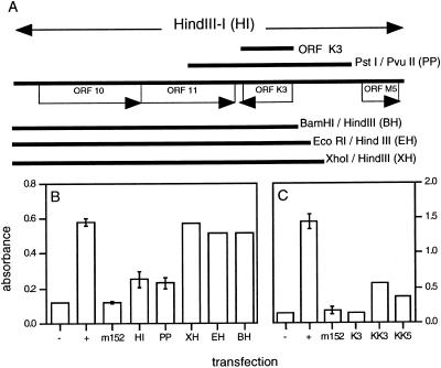
Figure 5.
Identification of a γHV-68 gene that blocks antigen presentation. (A) The HindIII-I fragment of γHV-68 is shown, together with the viral ORFs it contains (10) and the extents of the other viral fragments used for transfection. In each case, H-2Kb-SIINFEKL antigen presentation was assayed, using β-galactosidase production by the B3Z hybridoma. (B) L929-H-2Kb cells were transfected with pCDNA3-SIINFEKL polytope (1 μg), pCDNA3-ORF 50 (1 μg), and a genomic fragment as indicated (1 μg), either in pUC-119 or in pSV40-ZEO (Invitrogen). HI, PP, XH, EH, and BH refer to the γHV-68 genomic DNA fragments indicated in A. For “no epitope,” cells were transfected with pCDNA3-ORF 50 (1 μg) plus empty pCDNA3 vector (2 μg). For “no inhibitor,” cells were transfected with pCDNA3-polytope (1 μg), pCDNA3-ORF 50 (1 μg), and empty pCDNA3 vector (1 μg). As a positive control, cells were transfected with pCDNA3-polytope (1 μg), pCDNA3-ORF 50 (1 μg), and pCDNA3 containing the m152 inhibitory gene of murine cytomegalovirus (1 μg). (C) L929-H-2Kb cells were transfected with pCDNA3-polytope (1 μg) plus the K3 genomic ORF (K3) or the homologous KSHV K3 (KK3) or K5 (KK5) ORFs, each expressed in PCDNA3 (1 μg). Because all expression was driven by the cytomegalovirus immediate-early promoter in pCDNA3 for this experiment, pCDNA3-ORF 50 was not included in the transfection.

