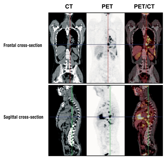
Figure 3.
Investigation of a patient with CUP syndrome using the PET/CT hybrid technique. The images are from a 42-year old male patient with a history of smoking, who first visited his GP with swelling of the cervical lymph nodes and left hematothorax. Extirpation of a cervical lymph node gave the histological diagnosis of squamous cell carcinoma. The tumor cells were TTF1-positive in immunohistology. The PET/CT investigation found multiple mediastinal lymph node metastases—right intraclavicular and paraaortic at the level of the celiac trunk—, also suspected pleural carcinosis. The present findings indicate a primary tumor in the left lung, even though this could not be directly identified. With the kind approval of Professor Uwe Haberkorn MD, Department of Nuclear Medicine, Heidelberg University

