Figure 2.
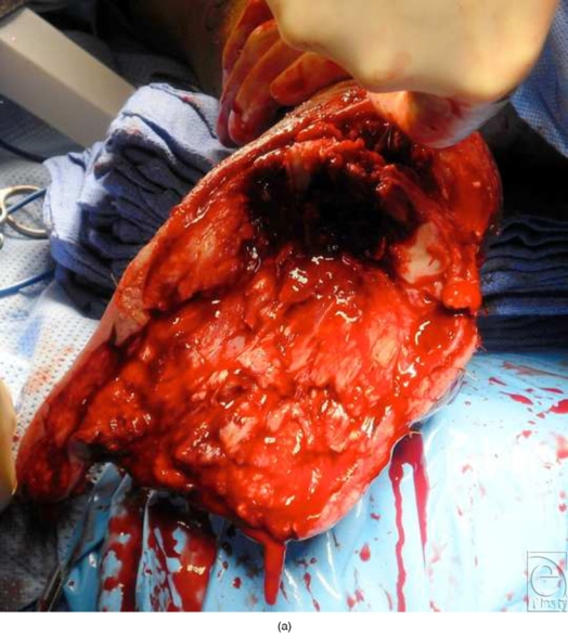
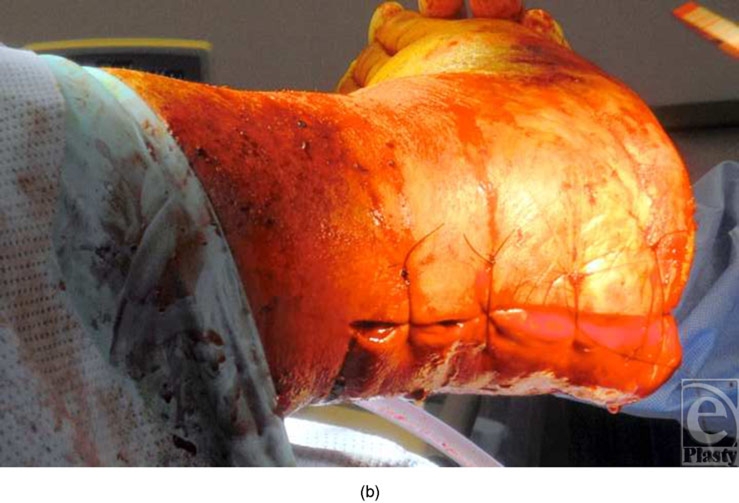
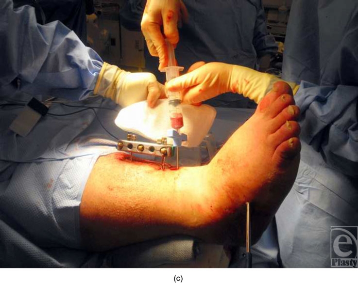
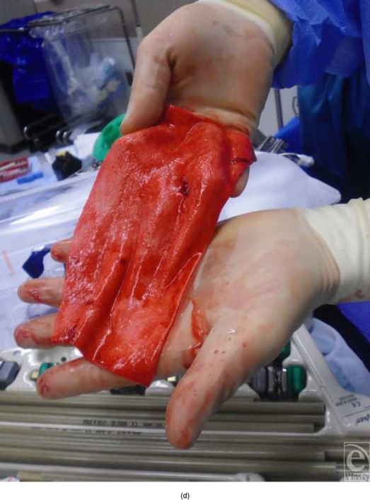
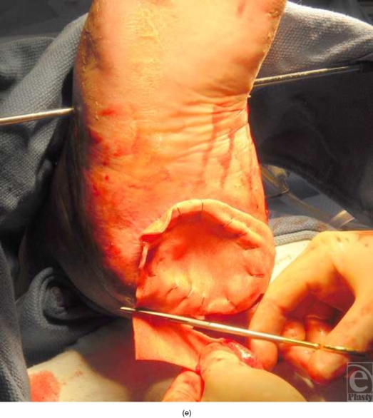
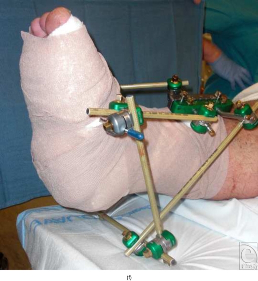
(a) Intraoperative photograph reveals a large, open, posterior heel wound after debridement. (b) Closure of posterior heel defect. (c) Bone marrow aspiration. (d) Preparation of acellular tissue matrix allograft. (e) Placement of acellular matrix. (f) SALSAstand construction around graft site demonstrating both posterior and plantar offloading of the posterior heel in addition to triplanar correction.
