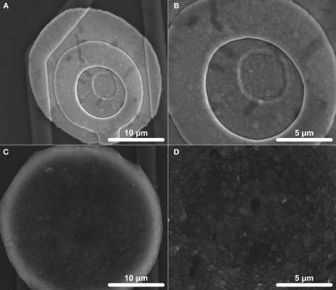
Figure 1.
Scanning electron microscopy (SEM) images of electrode surfaces (413 μm2). (A) An overall coating and (B) microscale surface morphology of IrOx which was activated by biasing bare Ir between +0.8 and −0.9 V at 1 Hz for 1600 s is shown. (C) An overall coating and (D) microscale surface morphology of PEDOT which was galvanostatically deposited at 6 nA for 900 s is shown.

