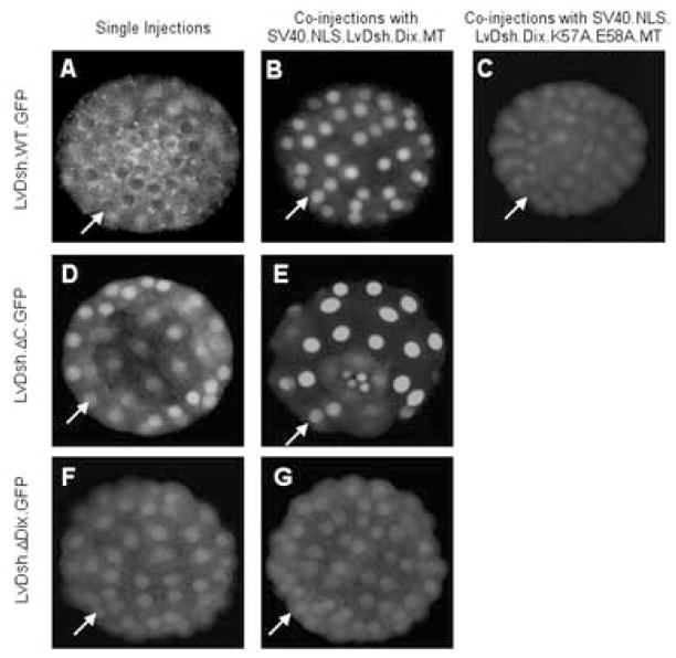
Figure 11.
LvDsh.DIX interacts via residues K57 and E58 with the DIX domain of full length LvDsh. LvDsh.GFP constructs were injected alone (A, D, and F) or co-injected with either nuclear-targeted LvDsh.DIX (B, E, and G) or nuclear-targeted mutant (K57A/E58A) LvDsh.DIX (C). Confocal images of representative embryos were taken at the blastula stage. Arrows indicate nuclei. When injected alone, wild-type LvDsh.GFP shows diffuse cytoplasmic staining with no nuclear accumulation (A). Co-injection with nuclear-targeted LvDsh.DIX results in a marked decline in cytoplasmic staining and a corresponding increase in nuclear LvDsh.GFP (B). The previously observed nuclear accumulation of LvDsh.ΔC.GFP is replicated when injected alone (D). Co-injection of LvDsh.ΔC.GFP with nuclear-targeted LvDsh.DIX greatly enhances the nuclear accumulation of the protein (E). The interaction between DIX and LvDsh is mediated by the DIX domain of the full-length protein, because LvDsh lacking the DIX domain (LvDsh.ΔDIX.GFP) shows minimal nuclear fluorescence in the absence (F) or presence (G) of nuclear-targeted LvDsh.DIX. Conserved, lipid-associating residues K57 and E58 within the DIX domain are required for its interaction with full length LvDsh.GFP (C).

