Figure 3.
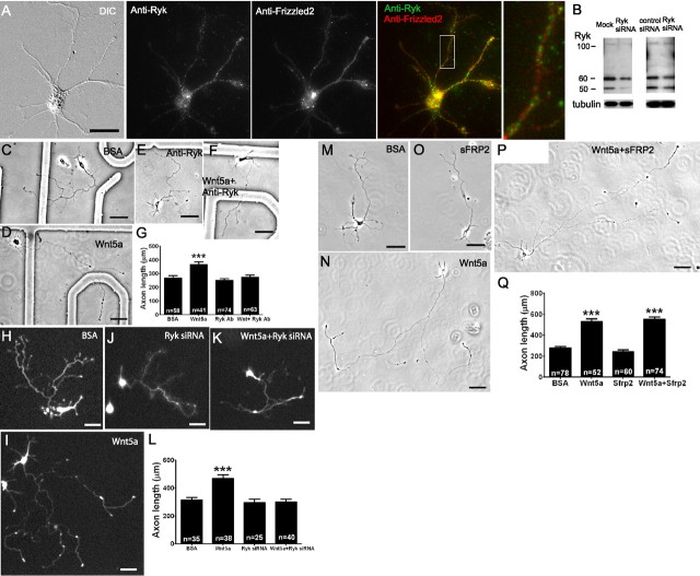
Ryk but not Fz receptors mediate Wnt5a-induced axon outgrowth. Immunocytochemistry with anti-Ryk and anti-Frizzled2 antibodies (A) show the presence of Ryk and Fz2 receptors on a pyramidal neuron from 2 d sensorimotor cortex cultured for 2–3 d before staining (scale bar, 20 μm). Left, A DIC image of the neuron stained for Ryk (green) and Fz2 (red) and the overlay showing both receptors are stained on the same neuron. Inset at right is from the box area at higher power. B, Western blot showing at left the presence of Ryk in P0 sensorimotor cortical neurons after 3–4 d in culture compared with neurons from the same tissue that were transfected with 100 pmol of Ryk siRNA. At right, the Western blot shows a decrease in Ryk expression following Ryk siRNA compared with a pool of nontargeting control siRNAs. C–F, Phase images of cortical neurons treated with BSA (control), Wnt5a, a function-blocking anti-Ryk antibody (10 μg/ml), or Wnt5a in the presence of anti-Ryk. H–K, Fluorescence images of cortical neurons transfected with GFP and treated with BSA (control) or Wnt5a, or transfected with GFP and Ryk siRNA and treated with BSA or Wnt5a. M–P, Phase images of cortical neurons treated with BSA, Wnt5a, 200 ng/ml sFRP2 (which blocks Wnt/Fz interaction), and sFRP2 in the presence of Wnt5a. Scale bar: C–Q, 50 μm. G, L, Q, Bar graphs of axon lengths of cortical neurons in conditions shown in C–F, H–K, and M–P, respectively. n, Numbers of axons in each condition in one experiment. Experiments were repeated three times. In all histograms, ***p < 0.001.

