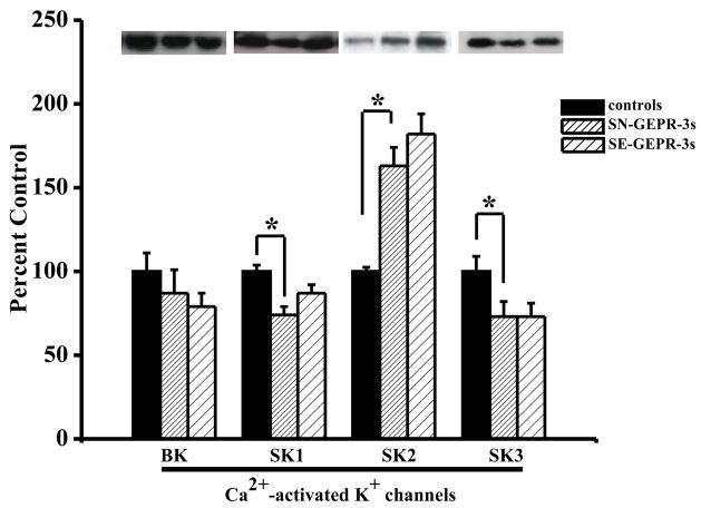
Figure 1.
Quantification of the protein expression of Ca2+-activated K+ channels. Representative immunoblots of BK, SK1, SK2 and SK3 subtypes of Ca2+-activated K+ channels obtained from IC neurons of control SD rats, SN-GEPR-3s and SE-GEPR-3s are showed in insets. The levels of protein expression of SK1 and SK3 subtypes of Ca2+-activated K+ channels were significantly decreased in IC neurons of SN-GEPR-3s compared to control SD rats. In contrast, the levels of proteins associated with SK2 channels were significantly increased in IC neurons SN-GEPR-3s compared to control SD rats. Seizure episodes slightly increased the protein expression of SK1 and SK2 channels in IC neurons compared to SN-GEPR-3s, but did not alter expression of SK3 channels. The levels of protein expression of BK channels were slightly reduced in SN-GEPR-3s compared to control SD rats as well as in SE-GEPR-3s compared to SN-GEPR-3s. Each column represents the mean ± S.E.M. (n = 8). *P <0.05 (ANOVA followed by post hoc test).

