
Figure 6.
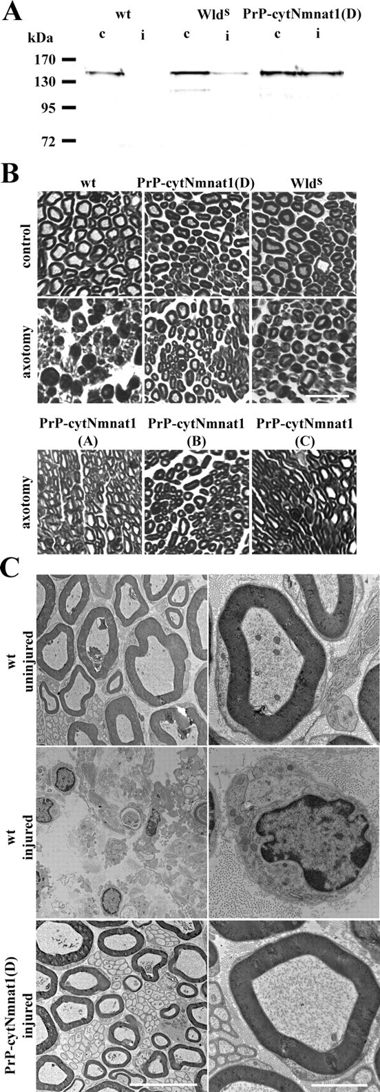
Axonal degeneration after sciatic nerve transection is delayed in PrP-cytNmnat1 Tg mice. A, Neurofilament (165 kDa component) levels remaining in the distal sciatic nerve segment 7 d after transection were measured by Western blot using anti-neurofilament antibody to examine axonal loss. Neurofilament levels in transected distal segment (i) of PrP-cytNmnat1(D) Tg mice were equivalent to those in the uninjured control (c) nerve. No neurofilament remained in the wild-type distal segment after injury. Transected distal segment from wlds mice also contained neurofilament. B, Plastic thin sections (4 μm) of distal sciatic nerve taken from PrP-Nmnat1(A–D) Tg, wlds, or wild-type mice 7 d after transection were stained with toluidine blue. Axons and the myelin structures of PrP-cytNmnat1 Tg and wlds mice were well preserved, whereas the wild-type nerve structure was completely disorganized with massive axonal loss and large amounts of myelin debris. C, Electron microscopic evaluation of these nerves revealed that the wild-type axonal structure was completely degenerated 7 d after axotomy (middle) compared with the uninjured nerve control (top). In contrast, the distal nerve from Prp-cytNmnat1(D) was well preserved (bottom). Higher magnification clearly showed an intact nerve structure including microtubules and mitochondria inside the axon in the transected transgenic nerve. Scale bars: B, 50 μm; C, left column, 10 μm; C, right column, 2 μm.