
Fig. 2. Photothrombosis causes neuronal death and astrocyte activation in the cortex.
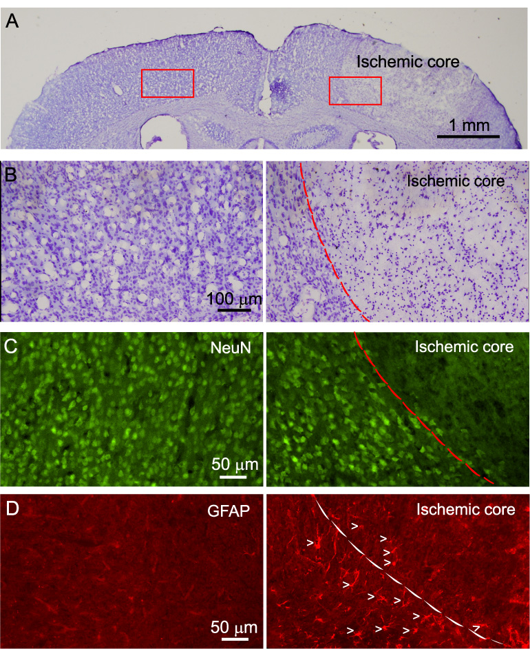
A) Nissl staining of brain sections showing cell damage in the ischemic region (right) 24 hr after photothrombosis. An area of 2 mm in diameter was illuminated in this experiment. B) High resolution micrographs of ischemic region (right) and corresponding region of contralateral side (left) from A. C) NeuN staining of a brain section showing neuronal death in the ischemic region (right) and healthy neurons in corresponding contralateral side (left). D) Enhanced GFAP expression in the transition zone between ischemic core and penumbra (right) after photothrombosis. The left panel shows low GFAP expression in the cortex in the contralateral side. Arrow heads indicate individual astrocytes. Dash line outlines the boundary between ischemic core and penumbral region.