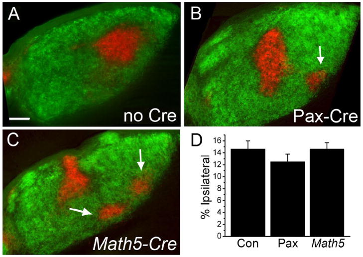
Figure 2.
Phr1 retinal mutants demonstrate defective localization of eye specific patches in the mature dLGN. (A) Following injection of labeled CTB into the ipsi- (red) and contralateral (green) eyes, control animals display a stereotyped pattern of innervation of the dLGN from each eye. (B,C) This pattern is disrupted in retinal Phr1 knockouts generated using the retina-specific Pax-Cre or the RGC-ubiquitous Math5-Cre lines and the Phr1 floxed allele. The ipsilateral patch is disrupted into multiple smaller patches (arrows). (D) The percentage of the nucleus occupied by ipsilaterally projecting axons is the same in controls (Con) and both Phr1 retinal mutants (Pax, Math5). Scale bar = 100 μm.

