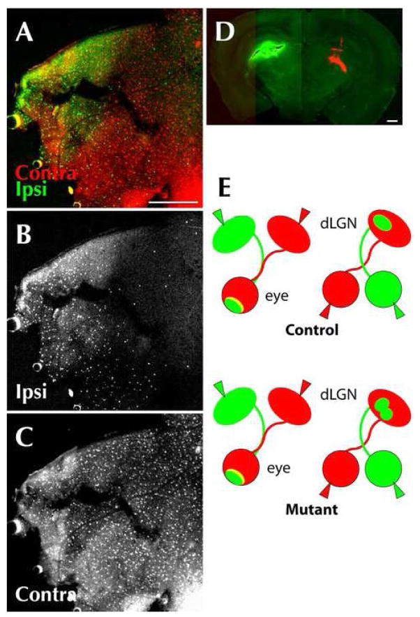
Figure 5.
RGCs from the inferotemporal retina innervate the ipsilateral thalamus in Phr1 retinal mutants. RGCs from the inferotemporal retina most often innervate the ipsilateral LGN, whereas RGCs from the remainder of the retina usually send axons across the optic chiasm and innervate the contralateral side. (A, B, C) Retrograde transport of CTB to the retina of αPax-Cre Phr1 mutant adult animals after injection of labeled choleratoxin. (D) Coronal section of the thalamus from the same animal showing conjugated CTB in the right (green) and left (red) thalamus. Flat mount of the retina of the right eye shows transport of CTB from the ipsilateral (green; A,B) and contralateral (red; A,C) thalamus of Phr1 retinal mutants. Note that despite a better fill of the CTB injection on the ipsilateral side (green), only the inferotemporal retina was labeled, unlike the contralateral injection (red) which resulted in labeling across the entire retina. This confirms that axons in the ipsilateral thalamus primarily originate from cell bodies in the inferotemporal retina, as expected. (E) Summary of eye and brain injection experiments: When the eyes of Phr1 retinal mutants are labeled (right) with two different colors, the pattern of label in the dLGN is altered compared to controls. However, when dLGNs are labeled (left), the pattern of label in the retinas is the same for mutants and controls, indicating that the result of the eye labeling experiment is not due to altered axonal crossing of the optic chiasm. Scale bars = 400 μm.

