
Figure 2.
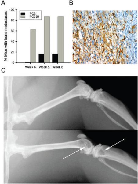
Characterization of a SCID mouse model of extravasation and bone metastasis. A, Comparison of extravasation ability of PC3 cells and PC3B1 cells. B, Immunohistochemistry analysis of α6 integrin expression on PC3 cells within the mouse trabecular bone using AA6NT antibody. C, Representative digital radiographs of mouse bone. Top panel displays normal bone, bottom panel indicates presence of osteolytic metastases in the distal femur and proximal tibia (arrows) at week 4.