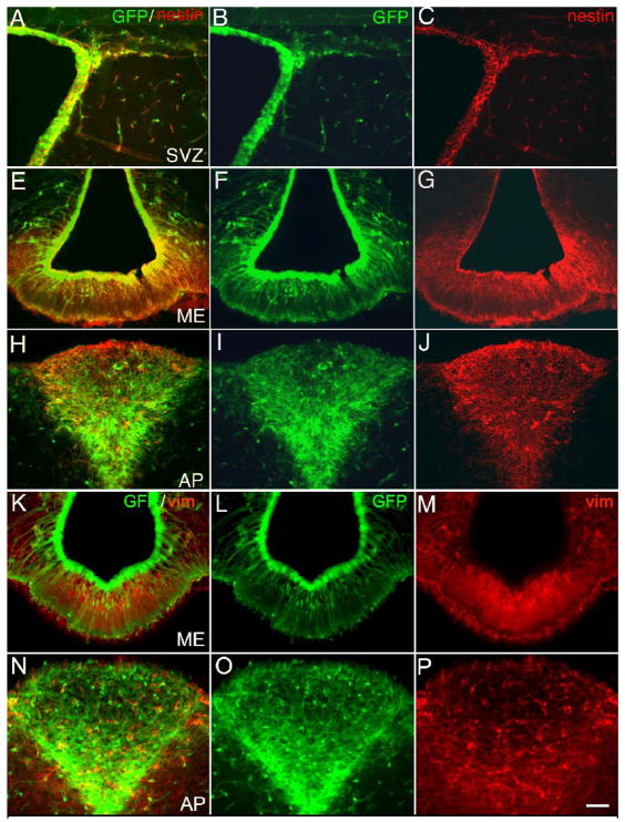
Figure 1.
Expression of intermediate filaments in CVOs of the adult transgenic nestin-GFP mouse. Using immunocytochemistry for tissue sections, cells in the SVZ (A-C), ME (E-G, K-M) or AP (H-J, N-P) were both GFP fluorescent (B, F, I, L, O) and nestin+ (C, G, J) or vimentin+ (M, P) in merged images (A, E, H, K, N). Calibration bar = 100μm.

