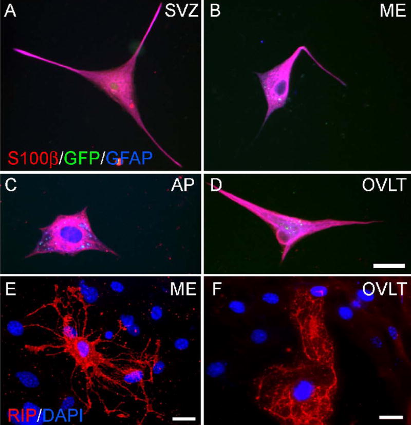
Figure 6.

Glial differentiation of nestin-GFP mouse cells in vitro. Undifferentiated nestin-GFP cells plated as adherent culture and differentiated for 9–14 days were analyzed by immunocytochemistry for expression of astrocyte markers S100β and GFAP and oligodendrocyte marker RIP. Shown are cells from the SVZ (A), ME (B), AP (C), and OVLT (D) co-expressing S100β and GFAP. Note that cells have lost expression of GFP, which would appear white if triple-labeled. ME (E) and OVLT (F) cells with characteristic oligodendrocyte morphology are shown expressing RIP. Calibration bar = 25 μm.
