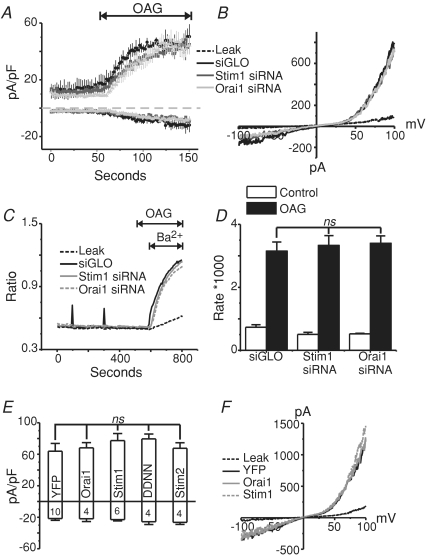
Figure 4. TRPC7 currents activated by OAG are not altered by the knockdown of STIM1 or Orai1, or by the over-expression of STIM1, STIM1 D76N/D78N, STIM2 or Orai1.
A, whole-cell OAG-activated currents (means ±s.e.m.) taken at −100 and +100 mV from stable TRPC7 cells transfected with siGLO (n= 3), STIM1 siRNA (n= 4) or Orai1 siRNA (n= 3) 72 h prior to the experiments. The external HBSS contained 2 mm Ca2+, and OAG (50 μm) was externally applied as indicated by the line above the graph. B, current–voltage (I–V) relationships taken from stable TRPC7 cells before (leak; broken black trace) and after (siGLO, STIM1 siRNA, Orai1 siRNA) focal OAG application. In all conditions, the outwardly rectifying I–V relationships were nearly identical. C, mean Ba2+ entries (fura-2 AM) in response to OAG (50 μm) application in stable TRPC7 expressing HEK293 cells 72 h after transfection with siGLO (Control), STIM1 or Orai1 siRNA. D, summary of data in C, showing no significant difference (ANOVA) of OAG-mediated Ba2+ influx in stable TRPC7 expressing cells expressing siGLO (n= 7 coverslips), STIM1 siRNA (n= 8 coverslips) or Orai1 siRNA (n= 4 coverslips). E, summary of OAG-mediated whole-cell currents (−100 mV and +100 mV) in stable TRPC7 cells transfected with eYFP (control) (n= 10), Orai1 (n= 4), STIM1 (n= 6), STIM1 D76N/D78N (n= 4) or STIM2 (n= 4). Nominally Ca2+-free HBSS was used as the external bathing solution in these studies. F, I–V relationship showing the currents recorded under the same conditions as E.

