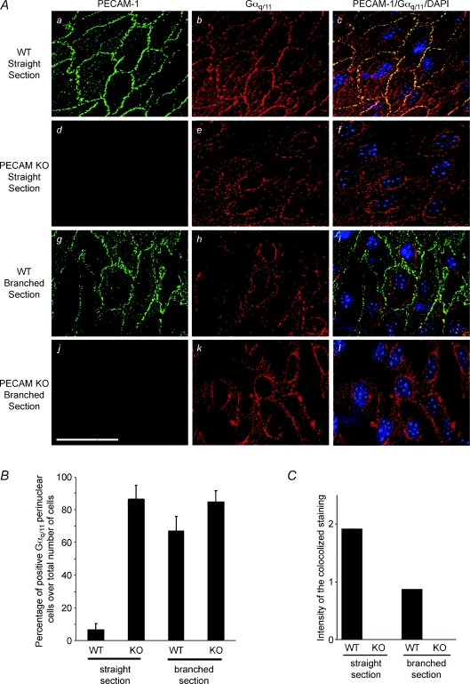
Figure 1. En face deconvolution microscopy of PECAM-1 and Gαq/11 staining in PECAM-1 knockout (KO) mouse aorta preparations.
A, staining was performed on the descending aorta (straight section) of wild-type (WT) mouse (a–c), PECAM-1 KO mouse (d–f) and branch points of the aorta (renal artery bifurcation) of WT (g–i) and KO mouse (j–l). Double staining was performed to localize PECAM-1 (green) and Gαq/11 (red). Superposition of PECAM-1 and Gαq/11 staining demonstrates co-localization in yellow–orange (right column). Cell nuclei were labelled using DAPI (blue). Scale bar is 20 μm. B, in WT descending aorta only a few cells expressed Gαq/11 staining in perinuclear areas. The number of cells with Gαq/11 perinuclear staining was significantly increased in PECAM-1-deficient mice and in branched regions of WT mice (n= 3 animals for each condition). C, the quantification of the Gαq/11 and PECAM-1 co-staining intensity (ranges of yellow) was reduced to half in branched section compared to straight section of the aorta. KO mice did not express PECAM-1.

