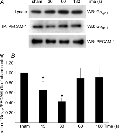
Figure 3. Time-dependent dissociation of Gαq/11–PECAM-1 complex in response to impulse flow.
Confluent serum-starved HUVECs were harvested 15, 30, 60 or 180 s after exposure to a single 1 s impulse (temporal gradient) using a syringe pump delivering a 14 dynes cm−2 FSS rate. A, representative Western blot (WB) of one experiment of sham and after 30, 60 and 180 s exposure to the impulse showing lysate Gαq/11, immunoprecipitated (IP) PECAM-1 blotted with Gαq/11 and PECAM-1. B, densitometric analysis of three independent experiments displays the kinetics of association–dissociation of the Gαq/11–PECAM-1 complex following impulse flow. Levels of Gαq/11 co-immunoprecipitated with PECAM-1 were normalized to lysate Gαq/11 levels and immunoprecipitated PECAM-1 levels. Results are percentage of sham controls. Values are mean ±s.d. (*P < 0.05 from sham control).

