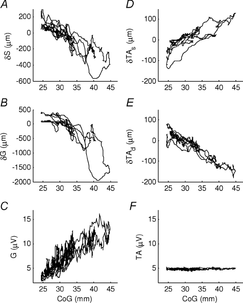
Figure 3. Muscular behaviour during normal standing.
Muscular change in muscle length and EMG during normal standing sways of representative subject (same trial as Fig. 2). The horizontal axis is the centre of gravity relative to the ankle joint centre position (CoG), 0 mm is the ankle joint centre position. The following panels show the muscular change in muscle length of A, soleus; B, gastrocnemius; D, superficial compartment of tibialis anterior; E, deep compartment of tibialis anterior. The following panels show the low-pass filtered superficial EMG of C, gastrocnemius and F, tibialis anterior. Negative displacement means shortening relative to the initial length. The two compartments of tibialis anterior show an opposite correlation (D vs. E) and the EMG does not appear to be modulated (F), but also soleus and gastrocnemius show different behaviour, in particular when the CoG is further backwards (A vs. B).

