Figure 2.
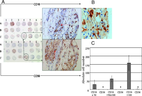
Renal cell carcinoma inflammatory infiltrates is mainly composed of CD16+ macrophages. (A) Two serial sections of RCC lesions (upper and lower panels) taken from the same areas of RCC specimens used for the construction of the indicated TAM. The punches in the upper panel were stained with a biotinylated anti-CD16 mAb, whereas those in the lower panel were stained with a biotinylated anti-CD56 mAb. Upper and lower right panels represent an enlargement of the indicated punches. Panel-positive cells are stained in brown. In either case, original magnifications of x20 are reported. (B) Detailed morphology of CD16+ cells identified in the indicated RCC area (original magnification, x60). A, right panel: Horizontal arrow indicates CD16+ interstitial cells, whereas vertical arrow indicates CD56+ RCC cells. (C) Absolute number of low levels of CD16+ cell infiltrate (≤50) and their relative CD56+ infiltrate, n = 48; intermediate levels of CD16+ cell infiltration and their relative CD56+ cell infiltration, n = 32; and high levels of CD16+ cell infiltration and their relative CD56+ cell infiltration, n = 28. Asterisks indicate CD56+ cell infiltrate.

