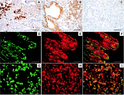
Figure 1.
Immunohistochemistry for IGF-IR in SPC-IGFIR (A), CCSP-IGFIR (B), or wild-type (C) mouse treated with doxycycline for 2 months. Immunofluorescence for IGF-IR (D) and CCSP (E) in a CCSP-IGFIR mouse and IGF-IR (G) and pro-SPC (H) in an SPC-IGFIR mouse treated with doxycycline. Merged images of IGF-IR/CCSP (F) and IGF-IR/SPC (I). Scale bars, 100 µm.

