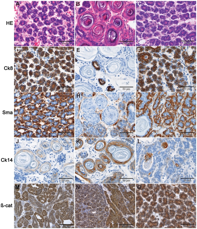
Figure 4. Differentiation.
HE staining of mammary tumors (A–B) and pulmonary metastases (C) from Apc +/1572T mice shows typical mammary glandular architecture and squamous differentiation. (D–F) Luminal epithelial differentiation as shown by cytokeratin 8 (Ck8) IHC staining. (G–I) Myoepithelial differentiation revealed by IHC staining with the Sma antibody. (J–L) IHC analysis with antibodies directed against cytokeratin 14 (Ck14) confirm the presence of squamous differentiation (hair follicle and skin cellular types). (M–O) β-catenin IHC analysis shows heterogeneous subcellular localization and intracellular accumulation with fewer cells characterized by positive nuclear staining. The results shown in this figure were confirmed in 12 independent primary tumors.

