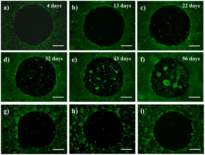
Figure 5.
Fluorescence micrographs of COS-7 cells seeded on a PEI/PVDMA film treated with a small drop of a solution of glucamine. Cells were allowed to grow continuously on the substrates for almost two months (see text for details). Images in (a–f) correspond to cells growing on the surface of this film (a) 4 days, (b) 13 days, (c) 22 days, (d) 32 days, (e) 43 days, and (f) 56 days after seeding. Cells were treated with Calcein AM prior to imaging. Images in (g–i) are fluorescence micrographs of COS-7 cells 72 hours after seeding on a glucamine-treated PEI/PVDMA film that was treated with trypsin and subsequently re-seeded with cells (g) one time, (h) two times, and (i) three times (see text). Scale bars = 500 μm.

