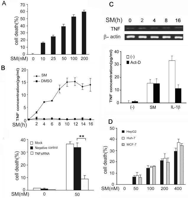
Figure 1. SMC3-induced transcription-independent TNF autocrine is required for SMC3-induced cytotoxicity in cancer cells.
A, H23 cells were treated with indicated concentrations of SMC3 for 36 h. Cell death was measured by LDH leakage assay. Data shown are the mean ± SD. B, H23 cells were treated with SMC3 (50 nM) or DMSO for the indicated times. The concentrations of TNF in cell culture media were measured by ELISA. C, TNF mRNA was detected by RT-PCR. β-actin was detected as an input control. D, H23 cells were pretreated with actinomycin D (10 μM) for 30 min followed by exposure to SMC3 (50 nM) or IL1β (5 ng/ml) for 8 h. TNF was detected as described in B. E, H23 cells were mock transfected or transfected with 5 nM of TNF-siRNA or negative control siRNA. Forty-eight hours after transfection, the cells were treated with SMC3 (50 nM) for 36 h or left untreated. The concentrations of TNF in cell culture media were measured As in A. **p < 0.01. F, H23 cells were pretreated with TNF neutralizing antibodies (1 μg/ml) or control antibody (1 μg/ml) for 1 h followed by SMC3 (50 nM) treatment for 36 h. Cell death was measured by LDH release assay. **p < 0.01. G, HepG2, Huh-7, and MCF-7 cells were treated with indicated concentration of SMC3 for 36 h. Cell death was measured by as described in A.

