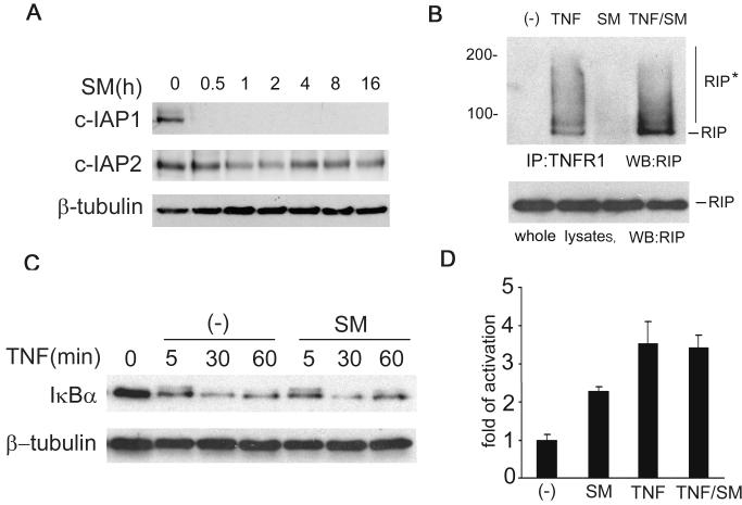
Figure 4. SMC3 does not inhibit TNF-induced NF-κB activation.
A, H23 cells were treated with SMC3 (50 nM) for the indicated times. cIAP-1 and cIAP-2 were detected by Western blot. β-Tubulin was detected as an input control. B, H23 cells were treated with SMC3 (50 nM) for 30 min followed by TNF (5 ng/ml) treatment for 10 min or left untreated. The cell lysates were immunoprecipitated with a TNFR1 antibody as described in Materials and Methods. RIP was detected by Western blot. The cell lysates (1% of the amounts used for IP) were probed for RIP as an input control. RIP*, modified RIP. C, H23 cells were treated with SMC3 (50 nM) for 30 min followed by TNF (5 ng/ml) for the indicated time points. IκBα was measured by We stern blot. β-Tubulin was detected as an input control. Note the phosphorylated IκBα shown as a weak shifted band at 5 min was comparable in samples treated with TNF alone or SMC3 plus TNF. D, H23 cells were cotransfected p5×κB-Luc and pRSV-LacZ. Twenty-four hours post-transfection the cells were treated as indicated. Luciferase activity was measured as in Fig. 2A.

