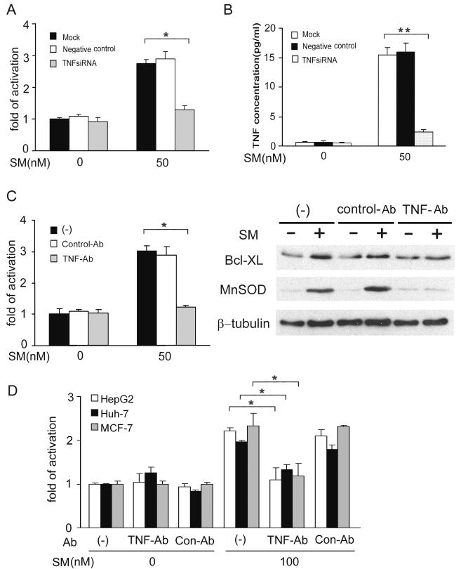
Fig 5. SMC3-induced NF-κB activation is TNF-dependent.
A, H23 cells were mock transfected or transfected with 5 nM of TNF-siRNA or negative control siRNA. Forty-eight h after transfection, the cells were cotransfected with p5×κB-Luc and pRSV-LacZ. After incubation for another 24 h, the cells were treated with SMC3 (50 nM) for 24 h or left untreated. Luciferase activity was detected and normalized to β-galactosidase activity. *p < 0.05. B, H23 cells were treated as in A. The concentration of TNF in culture media was determined by ELISA. **p < 0.01. C, H23 cells were cotransfected p5×κB-Luc and pRSV-LacZ. Twenty-four hours post-transfection the cells were pretreated with TNF neutralizing antibodies (1 μg/ml) or control antibody (1 μg/ml) for 1 h followed by 50 nM SMC3 treatment for 24 h. Luciferase activity was detected as in A. *p < 0.05. D, H23 cells were pretreated with TNF neutralizing antibodies (1 μg/ml) or control antibody (1 μg/ml) for 1 h followed by 50 nM SMC3 treatment for 24 h or left untreated. Bcl-XL and MnSOD were detected by Western blot. β-Tubulin was detected as an input control. E, HepG2, Huh-7, and MCF-7 cells were transfected, treated, and analyzed as described in C. *p < 0.05.

