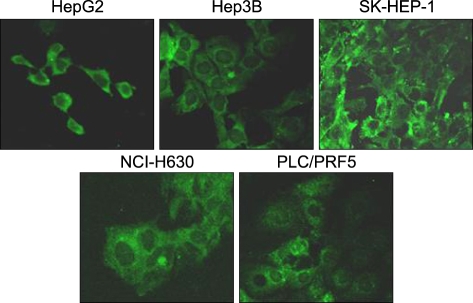
Fig. 3.
Immunofluorescence staining for CXCR4 in the liver cancer cell lines. The cells were incubated with 12G5, reacted with a secondary antibody labeled with FITC and then they were subjected to confocal microscopic examination. Note the abundant cytoplasmic expression of CXCR4 in all of the cells.

