Figure 2.
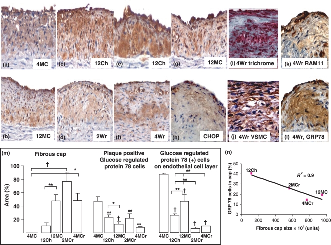
Photomicrographs of glucose regulated protein 78 (GRP78) positive cells in the initiation, progression and regression of atherosclerosis. GRP78 positive cells, most probably macrophages, are present in high amounts in aortic plaques in 4MC (a). GRP78 positive cells remained throughout the duration of 12 week dietary manipulation (12Ch – c and e; 12MC, g and b) and during the regression period (2Wr – d and 4Wr – f). CHOP immunoreactivity was also present in cells overlying endothelium and not in core (h). The trichrome stain (i) reveals collagenous cap forming (blue) with other cells (red), which are most probably VSMC (j) and some macrophages (k). Serial sections (K, RAM11 and L, GRP78) show that GRP78 positive cells are likely to be macrophages. GRP78 positive cells within plaques significantly decreased from 4 weeks onwards (m), and GRP78 positive cells overlying endothelia was scarce in the regression groups (m). Quantification of percentage area of GRP78 positive cells in the fibrous cap showed a strong correlation between fibrous cap size and cells in the cap (N, r2 = 0.9). Quantification of fibrous cap showed a significant increase in fibrous cap content in 12MC compared with 12Ch, and the regression groups also showed marked fibrous cap development (l). Results are expressed as mean ± SEM, *P < 0.05, †P < 0.01, ‡P < 0.001.

