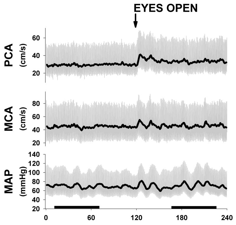
Figure 1.
Example of one subject during eyes closed and open protocol. Subjects had eyes closed for 2 min and then were instructed to open eyes and were monitored for another 2 min. Note clear activation in PCA after eyes opening with no change in MCA velocities. Dark bars represent one min steady state sections used for transfer function analysis.

