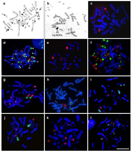
Fig. 3.
C-banding, Ag-NOR staining, and FISH examples. (a) C-banded BOE metaphase (the arrow shows the W chromosome). (b) Ag-NOR staining of BOE chromosomes (the arrow indicates the association of the two chromosomes with NORs). (c) The probe from a Chinese pangolin (Manis pentadactyla) BAC clone containing ribosomal DNA hybridized to BOE chromosome 13. (d) BOE 15+16 (red) and 17+18+19+20 (green) probes hybridized to BOE metaphase; note that the latter also gave cross-hybridization signals to BOE 13p proximal and W (see Fig. 1d and e for details); arrows show BOE 17–20. (e) BOE 7 (red) probe hybridized to BOE metaphase. (f) GGA probes R7 (green) and R9 (red) hybridized to GGA metaphase. (g) GGA 9 (green), R3 (red) and R6 (pink) probes hybridized to BOE chromosomes 7, 13q and 16q. (h) GGA probes R7 (green) and R9 (red) hybridized to BOE chromosomes 11, 13, 15, 17–20; (i) GGA probes 4 (green) and 9 (red) hybridized to BOE chromosomes 4, 7 and 8. (j) BOE probes 6 (green) and 10 (red) hybridized to GGA chromosomes 5 and two pairs of microchromosomes. (k) BOE probe 7 hybridized to GGA chromosomes 9 and two pairs of microchromosomes. (l) BOE probes 15+16 (green) and 17+18+19+20 (red) hybridized to 4 pairs of GGA microchromosomes. Scale bar represents 10 μm

