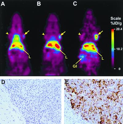
Figure 4.
Serial microPET scans of a mouse bearing bilateral C6 (arrowhead) and LS174T (arrow) xenografts, injected with 64Cu minibody and imaged at 2 h (A), 6 h (B), and 24 h (C). After the final scan, tumors were excised and subjected to immunohistochemical staining by using anti-CEA cT84.66 as the primary antibody. Photomicrographs of C6 (D) and LS174T (E) xenografts.

