FIG. 2.
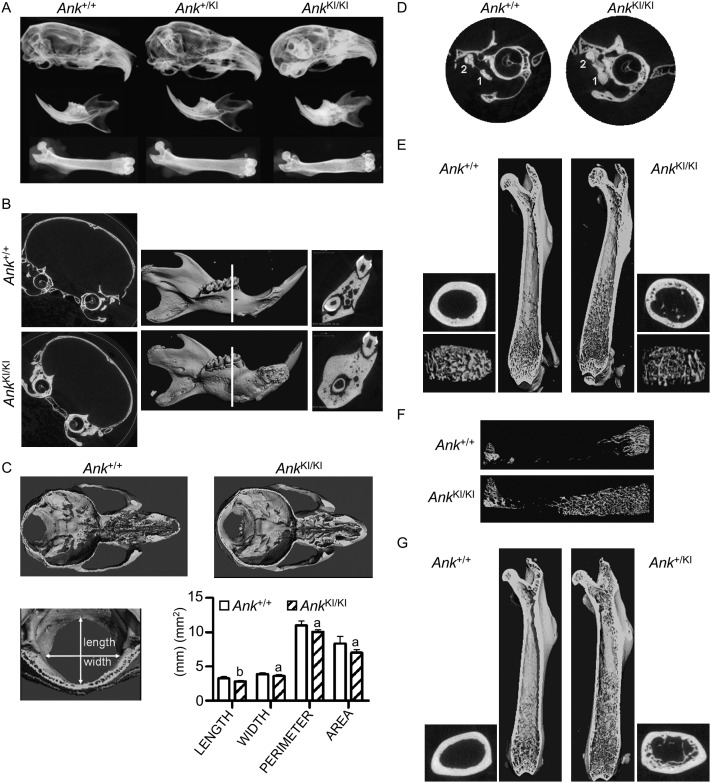
CMD-like phenotype in Ank KI/KI mice. (A) Representative radiographs of skulls, mandibles, and femurs from 6-mo-old Ank +/+, Ank +/KI, and Ank KI/KI male mice. (B) μCT images of frontal plane through skulls and 3D reconstruction of mandibles from 3-mo-old Ank +/+ and Ank KI/KI male mice. White line indicates sagittal plane through furcation of first mandibular molar. (C) Internal, dorsal view of cranial floor and nasal cavities from horizontal plane of superior semicircular duct and the cribriform plate of ethmoid. Histogram shows dimensions of foramen magnum of Ank +/+ and Ank KI/KI littermates. (D) 2D μCT images of tympanic bulla from frontal plane through cochlea showing fusion of malleus (1) and incus (2). (E) Internal view of Ank +/+ and Ank KI/KI femurs. 3D reconstructions of trabeculation in metaphysis and cross-sectional slices of cortical bone in diaphysis. (F) 3D μCT images of total trabecular bone in femurs from Ank +/+ and Ank KI/KI mice at 10 wk of age. (G) Internal view of femurs of 12-mo-old Ank +/+ and Ank +/KI mice. 3D reconstructions of trabeculation in metaphysis and cross-sectional slices of cortical bone in diaphysis.

